
Twist-on efocus Fluorescence Microscopy System (OBSOLETE)
Twist-on efocus Fluorescence Microscopy System (OBSOLETE)
Voici une description courte popur vouir ce que ca donne seulement... Lorem ipsum dolor sit amet, consectetur adipiscing elit, sed do eiusmod tempor
…generating
The Twist-on efocus Fluorescence Microscope enables users to visualize larger brain areas in freely behaving animals studies. The large field of view of up to 650 x 650 microns and the electronic depth adjustment of 300 microns allows a larger volume to perform calcium imaging at cellular resolution. This microscope offers an optimized way to attach the microscope body to the imaging cannula with precision, with a simple barrel. The attachment/detachment is now easier and does not require any tools.
Motor assisted rotary joint (AHRJ) and dummy microscope bodies are available on option.
Motor assisted rotary joint (AHRJ) and dummy microscope bodies are available on option.
The GCaMP Twist-on efocus microscopy system is optimized for GFP-like fluorescence proteins imaging.
The system includes:

- Twist-on efocus Green Fluorescence Microscope Body, the head mounted microscope
- Fluorescence Microscope Driver, to control the microscope
- Electrical cable for efocus Fluorescence Microscope Bodies, to connect the microscope to the driver
- Connectorized LED
- Optical fiber patch cords, to connect the light source to the microscope
- Fluorescence Microscope Holder 400
- Clamp for Fluorescence Microscope Holder
- Doric Neuroscience Studio Software
Add optional items:
Complete your system with:
| Field of View | 650 µm x 650 µm |
| Working distance adjustment range | 0 - 300 µm |
| Frame rate | up to 45 fps |
| Fluorescence recording | |
|---|---|
| Excitation | 458 / 35 nm |
| Emission | 525 / 40 nm |
- The Twist-on efocus Fluorescence Microscope enables users to visualize larger brain areas in freely behaving animals studies. The large field of view of up to 650 x 650 microns and the electronic depth adjustment of 300 microns allows a larger volume to perform calcium imaging at cellular resolution. This microscope offers an optimized way to attach the microscope body to the imaging cannula with precision, with a simple barrel. The attachment/detachment is now easier and does not require any tools.
Motor assisted rotary joint (AHRJ) and dummy microscope bodies are available on option. -
The GCaMP Twist-on efocus microscopy system is optimized for GFP-like fluorescence proteins imaging.
The system includes:
- Twist-on efocus Green Fluorescence Microscope Body, the head mounted microscope
- Fluorescence Microscope Driver, to control the microscope
- Electrical cable for efocus Fluorescence Microscope Bodies, to connect the microscope to the driver
- Connectorized LED
- Optical fiber patch cords, to connect the light source to the microscope
- Fluorescence Microscope Holder 400
- Clamp for Fluorescence Microscope Holder
- Doric Neuroscience Studio Software
Add optional items:
Complete your system with:
Field of View 650 µm x 650 µm Working distance adjustment range 0 - 300 µm Frame rate up to 45 fps Fluorescence recording Excitation 458 / 35 nm Emission 525 / 40 nm - The RCaMP Twist-on efocus microscopy system is optimized for Red Fluorescence Proteins imaging.
The system includes:
- Twist-on efocus Red Fluorescence Microscope Body, the head mounted microscope
- Fluorescence Microscope Driver, to control the microscope
- Electrical cable for efocus Fluorescence Microscope Bodies, to connect the microscope to the driver
- Ce:YAG Light Source and its driver with 540/15 nm bandpass filter for Red Fluoescent Proteins excitation
- Optical fiber patch cords, to connect the light source to the microscope
- Fluorescence Microscope Holder 400
- Clamp for Fluorescence Microscope Holder
- Doric Neuroscience Studio Software
Add optional items:
Complete your system with:
Field of View 650 µm x 650 µm Working distance adjustment range 0 - 300 µm Frame rate up to 45 fps Fluorescence recording Excitation 540 / 15 nm Emission 609 / 57 nm - The GCaMP + Red Opsins Twist-on efocus microscopy system is optimized for GFP-like fluorescence proteins imaging and Red Opsins (e.g. NpHR3.0, Chrimson, ...) activation.
The system includes: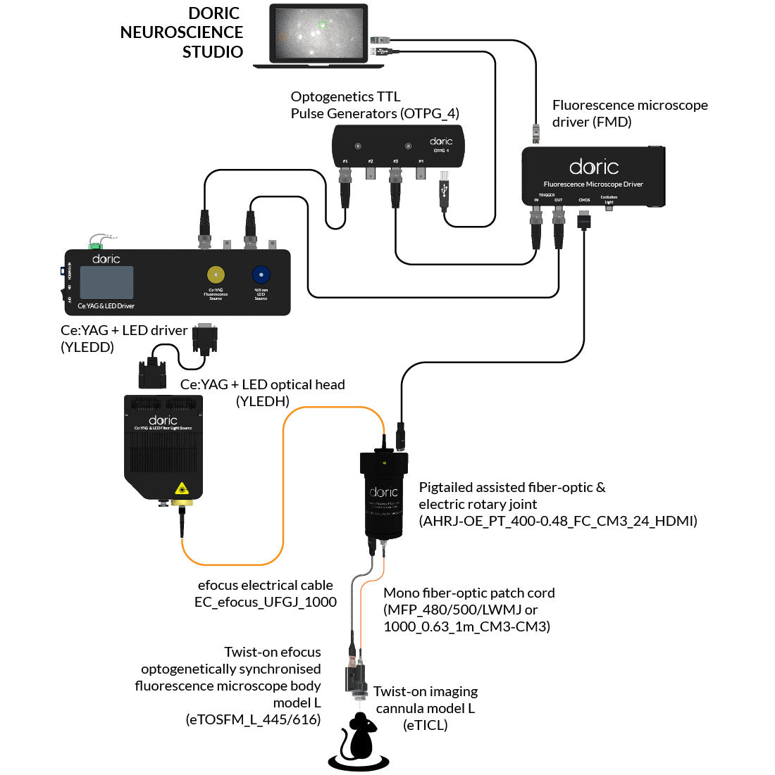
- Twist-on efocus GCaMP/NpHR Fluorescence Microscope Body, the head mounted microscope
- Fluorescence Microscope Driver, to control the microscope
- Electrical cable for efocus Fluorescence Microscope Bodies, to connect the microscope to the driver
- Ce:YAG + LED Light Source and its driver with 612/69 nm bandpass filter, for GCaMP excitation and Red Opsins activation
- Optogenetics TTL Pulse Generators 4 Channels, for imaging and optogenetic stimulation synchronization
- Optical fiber patch cords, to connect the light source to the microscope
- Fluorescence Microscope Holder 400
- Clamp for Fluorescence Microscope Holder
- Doric Neuroscience Studio Software
Add optional items:
Complete your system with:
Field of View 650 µm x 650 µm Working distance adjustment range 0 - 300 µm Frame rate up to 45 fps Fluorescence recording Excitation 458 / 35 nm Emission 525 / 40 nm Optogenetic stimulation Wavelength 612 / 69 nm Activation intensity 0 to 55 mW / mm2 - The Red Fluorescence + Blue Opsins Twist-on efocus microscopy system is optimized for Red Fluorescence Proteins imaging and Blue Opsins (ChR2) activation.
The system includes: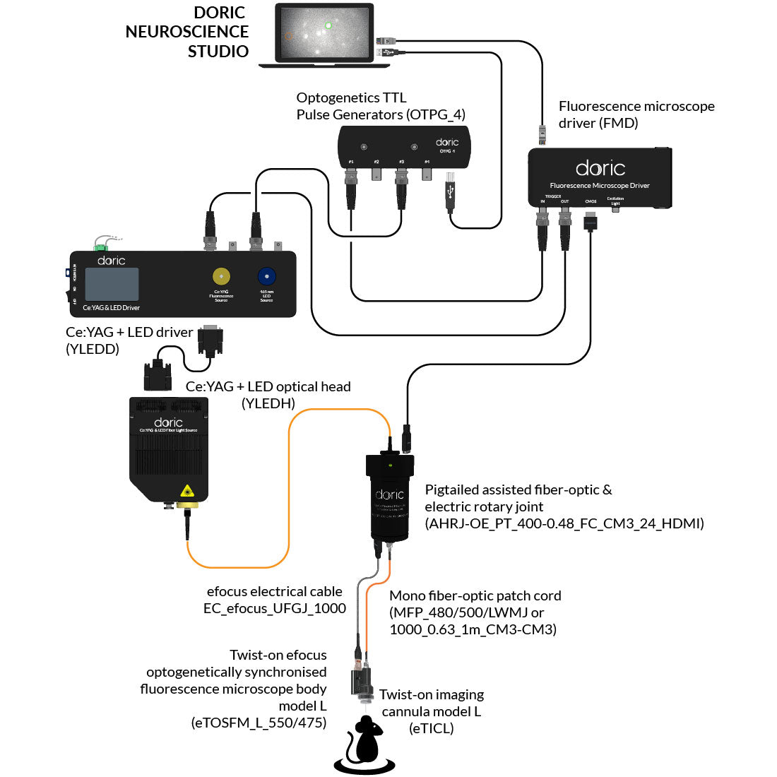
- Twist-on efocus RCaMP/CHR2 Fluorescence Microscope Body
- Fluorescence Microscope Driver, to control the microscope
- Electrical cable for efocus Fluorescence Microscope Bodies, to connect the microscope to the driver
- Ce:YAG + LED Light Source and its driver with 540/15 nm bandpass filter, for Red Fluorescence Proteins excitation and ChR2 activation
- Optogenetics TTL Pulse Generators 4 Channels, for imaging and optogenetic stimulation synchronization
- Optical fiber patch cords, to connect the light source to the microscope
- Fluorescence Microscope Holder 400
- Clamp for Fluorescence Microscope Holder
- Doric Neuroscience Studio Software
Add optional items:
Complete your system with:
Field of View 650 µm x 650 µm Working distance adjustment range 0 - 300 µm Frame rate up to 45 fps Fluorescence recording Excitation 540 / 15 nm Emission 609 / 57 nm Optogenetic stimulation Wavelength 465 / 20 nm